When an ankle sprain needs a second look
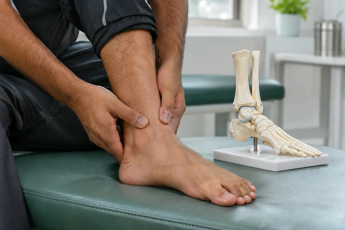
The bruising has faded and the swelling is smaller, but your ankle still aches deep inside when you walk, run or use stairs. After a sprain, it is easy to assume that the ligaments simply need more time. Sometimes they do. However, persistent joint pain can also come from damage to the cartilage and bone beneath it, called an osteochondral lesion of the talus, or OLT.
This possibility does not mean every slow recovery needs surgery. It means the cause of continuing symptoms deserves assessment rather than repeatedly testing the ankle through painful exercise.
What is an osteochondral lesion of the talus?
The talus sits between the lower-leg bones and the heel, forming the lower part of the ankle joint. Its upper surface is covered by smooth cartilage that helps the joint move. An osteochondral lesion affects this cartilage and the underlying bone. The injury can range from a small damaged area to a cyst beneath the surface or a partly detached fragment.
During an ankle twist, the talar surface can strike another part of the joint. Ligaments may be injured at the same time. Some lesions develop without a clearly remembered accident, so the absence of a dramatic injury does not rule them out.
Which symptoms may suggest more than a sprain?
Possible symptoms include deep ankle pain with weight-bearing, swelling that returns after activity, and difficulty progressing back to sport. Some people notice catching, locking or a feeling that movement is obstructed. Pain may be hard to locate precisely.
These symptoms are not specific to an OLT. Tendon injuries, ligament instability, impingement and an unrecognised fracture can also cause ongoing problems. A lesion can be present without locking, while painless clicking alone does not establish cartilage damage. What matters is the pattern, its effect on function and whether recovery is progressing.
How is this different from ankle instability?
Instability often involves repeated rolling or giving way, whereas an osteochondral lesion involves the joint surface. The two can coexist. Your clinician therefore assesses stability as well as tenderness, swelling, range of movement and walking pattern.
The article on ankle instability after repeated sprains explains the ligament side of this problem. Treating a scan finding in isolation can overlook another contributor to pain or repeated injury.
Can an X-ray miss a talar cartilage injury?
Yes. X-rays help assess alignment, fractures and visible changes in the bone, but some osteochondral lesions are not apparent on the first images. A normal X-ray does not directly demonstrate that all the cartilage is healthy.
When an OLT is clinically suspected, MRI can assess cartilage, underlying bone and surrounding soft tissues. CT may be useful for defining bone defects or cysts and planning a procedure. These tests answer different questions; not everyone needs both. MRI findings also need interpretation because swelling within bone can affect how large a lesion appears.
Bring previous images, not just reports, and explain what treatment you have already tried. Ask whether further imaging would change the plan rather than assuming a more detailed scan automatically means an operation.
What are the non-surgical treatment options?
Selected stable lesions can be managed without surgery. Care may include activity modification, a period of protection or restricted weight-bearing, suitable pain relief and rehabilitation. A clinician should decide whether a brace, boot or walking aid is appropriate and when loading can increase.
Physiotherapy is adapted to symptoms and the lesion, rather than following the same exercises for every sprain. Improvement is assessed through pain, swelling and function, with follow-up imaging when needed. Symptoms and scan appearances do not always change together. There is no single recovery timetable that fits all lesions.
When might surgery be considered?
A displaced or unstable fragment, significant mechanical symptoms, or persistent pain despite appropriate non-operative treatment may lead to a surgical discussion. Lesion size, depth, location, cartilage condition and previous treatment influence the choice.
Options can include arthroscopic assessment and treatment, fixation of a suitable fragment, bone-marrow stimulation, or cartilage-and-bone restoration procedures. These are not interchangeable, and none guarantees a particular return-to-sport date. Our ankle injury assessment and treatment page outlines the broader evaluation pathway. Associated instability may also need attention when planning care.
When should ankle pain be checked urgently?
Seek prompt care for worsening severe pain, major swelling, inability to bear weight, or fever with an injured ankle. A deformed ankle, new numbness, or a cold or discoloured foot needs emergency assessment. Do not wait for a routine appointment when these warning signs occur.
If deep pain or recurrent swelling continues after a sprain, book an orthopaedic consultation with Dr Rahul Grover in Delhi. A careful review can distinguish ligament recovery from joint-surface injury and guide the next step without assuming that every persistent symptom needs surgery.
Educational references: AOFAS FootCareMD, Osteochondral Lesions; Walther et al., DGOU recommendations (2023); The role of imaging in osteochondral lesions of the talus, British Journal of Radiology (2024); Steele et al., Current Concepts in Diagnosis and Treatment (2023 republication); NHS, Sprains and strains. General information only, not a substitute for individual medical advice.