Why a normal X-ray may not explain new knee pain
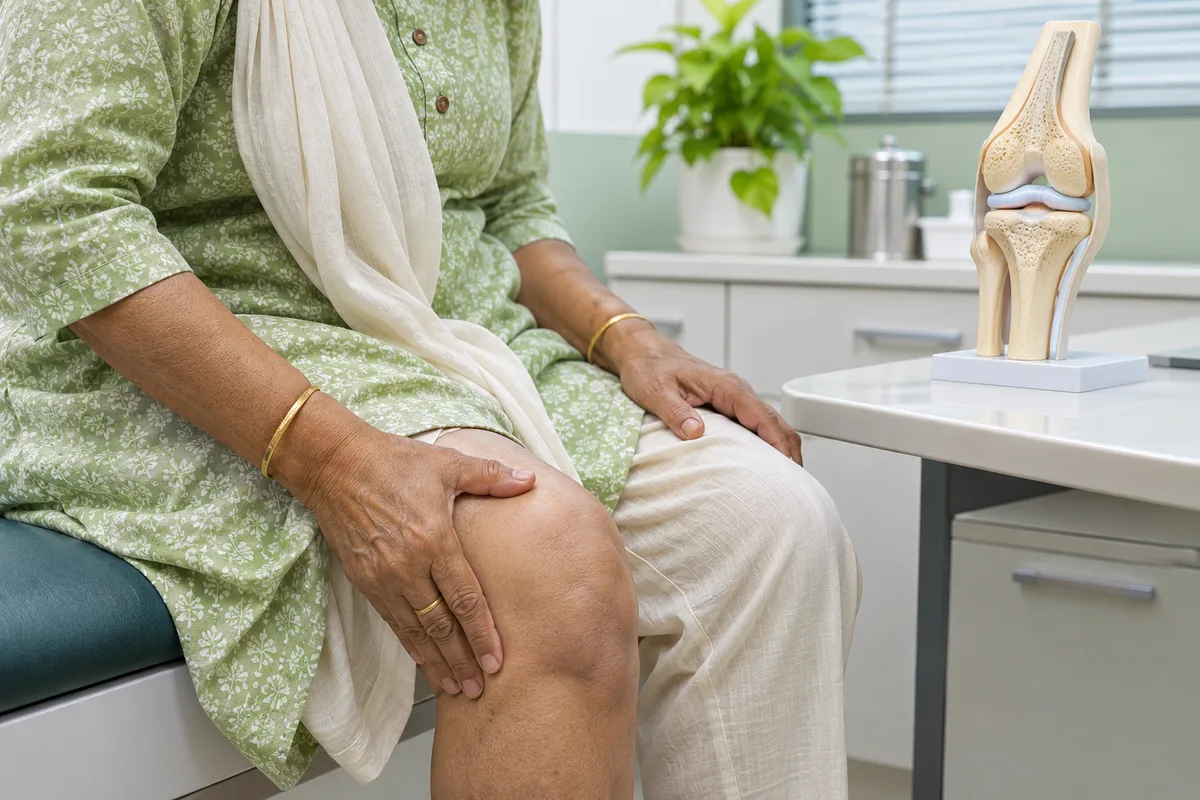
Sudden knee pain without a fall or sporting injury can be confusing. You may remember the day walking became painful, yet your X-ray shows little more than mild wear. One possible explanation is a subchondral insufficiency fracture of the knee, often shortened to SIFK. It is not the cause of every unexplained knee ache, but recognising it matters because the initial treatment can differ from routine arthritis rehabilitation.
What is a subchondral insufficiency fracture?
Subchondral means beneath the joint cartilage. An insufficiency fracture develops when the bone in this area cannot tolerate the load placed on it. Unlike a fracture caused by a major accident, it can occur during ordinary activity. The weight-bearing inner part of the thigh bone at the knee is a common location.
SIFK is seen particularly in middle-aged and older adults. Reduced bone strength may contribute, but osteoporosis is not present in every case. Changes in how the knee distributes pressure also matter. This is why simply taking calcium, or assuming age-related wear is the whole explanation, may miss the underlying problem.
Which symptoms deserve closer review?
The pain may begin suddenly or increase over a short period. Standing and walking often aggravate it, and there may be tenderness over a specific part of the knee. Some people notice swelling or pain at rest and at night. These features overlap with other conditions and cannot confirm a fracture on their own.
Tell your clinician whether the discomfort is new, different from previous arthritis pain, or severe enough to change your walking. Mention recent increases in activity, previous knee surgery, and any difficulty bearing weight. A clear history is often more useful than describing the scan as simply normal or abnormal.
Why can the first X-ray look normal?
X-rays are useful for examining joint alignment, arthritis and established bone damage. However, an early insufficiency fracture may be too subtle to show. Changes such as flattening or collapse of the bone surface can become visible later. A reassuring initial X-ray therefore does not exclude every important cause of pain.
MRI can reveal a fracture-related line and changes in the surrounding bone marrow before clear X-ray abnormalities develop. It also shows cartilage and meniscal structures. MRI is not necessary for every sore knee: it is considered when the examination and symptom pattern raise concern or when persistent pain is not explained by initial imaging.
How are meniscus injuries connected?
The meniscus helps distribute forces across the knee. Certain tears, particularly root tears, or displacement of the meniscus can change that load distribution. Meniscal problems and SIFK may coexist, so identifying a tear does not necessarily explain the entire pain picture.
Our article on meniscus tears, clicking and knee swelling explains why examination and imaging need to be considered together. Treatment should address the clinically important findings rather than just the most familiar term on the report.
Does a hidden knee fracture always need surgery?
No. Early lesions without collapse may be managed with protected weight-bearing, appropriate pain relief and monitored recovery. Your clinician may recommend walking aids and explain how much load is suitable. Do not push through painful running, long walks or heavy squats while the cause is being assessed.
The duration of protection and the exercise programme must be individualised. Research has not established one rehabilitation schedule suitable for everyone. Follow-up considers symptoms and, where appropriate, repeat imaging. Recovery can be prolonged, so a fixed promise of returning to normal within a few weeks is not reliable.
If the bone surface collapses, symptoms persist or arthritis is advanced, surgical options may be discussed. The choice depends on the location and extent of damage, the rest of the joint and your health. Our fracture assessment and treatment page describes the broader approach to deciding between protection and surgery; not every fracture needs fixation.
When should you seek urgent help?
Seek urgent medical assessment if you cannot bear weight, cannot move the knee, have severe pain or major swelling, or the knee changes shape. A hot, red knee with fever or feeling unwell needs prompt assessment for infection or another urgent cause. Do not wait for a routine MRI appointment in these circumstances.
For ongoing symptoms, book an orthopaedic consultation with Dr Rahul Grover in Delhi and bring the actual scans, reports and previous treatment details. Ask whether your symptoms fit the X-ray findings and whether further imaging would change the plan. The aim is to identify the cause early without assuming that every painful knee needs an operation.
Educational references: Ochi et al., review of SIFK imaging and differential diagnoses (2022); Zimmerman et al., Perspective on subchondral insufficiency fracture of the knee (2021); Malghem et al., subchondral bone lesions and imaging (2023); Subchondral Insufficiency Fractures of the Knee: A Clinical Narrative Review (2023); NHS, Knee pain. General information only; this article does not replace individual medical advice.