Understanding a knee that will not fully straighten
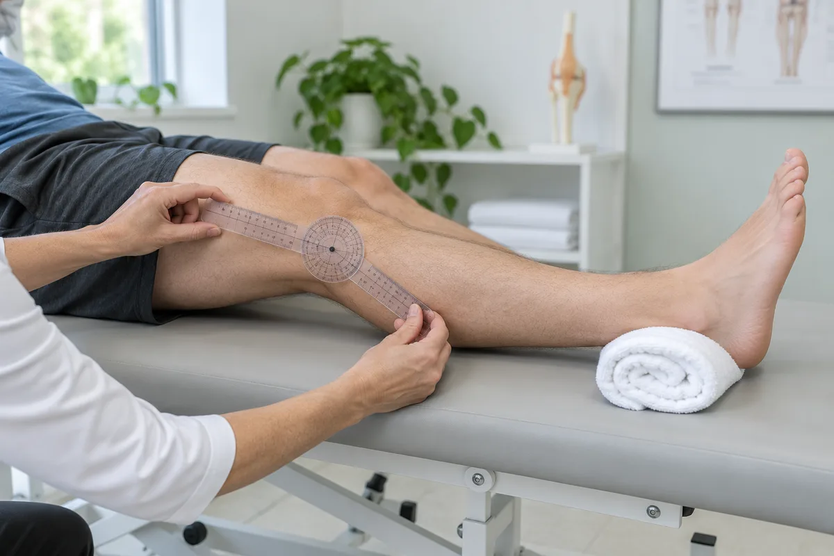
After ACL reconstruction, people often focus on bending the knee, climbing stairs and rebuilding strength. Straightening the knee fully matters too. If the final part of extension remains restricted, walking can feel awkward even when the knee feels stable. Persistent stiffness deserves an explanation, not simply a harder exercise programme.
One possible cause is a cyclops lesion, a small nodule of scar tissue near the reconstructed ligament. However, not every stiff knee has this problem, and finding a lesion on MRI does not automatically mean another operation is needed.
What is a cyclops lesion after ACL reconstruction?
A cyclops lesion is a localised collection of fibrous tissue, usually in front of the ACL graft within the knee. In some patients, it catches between structures as the knee approaches full extension. The combination of this tissue and a symptomatic mechanical block is called cyclops syndrome.
The original clinical description of cyclops syndrome reported loss of extension after ACL reconstruction and improvement following arthroscopic treatment. This is different from saying that every postoperative scar or click needs removal. The examination must show how the finding relates to the person's symptoms.
How is it different from ordinary postoperative stiffness?
Swelling, discomfort and difficulty activating the thigh muscles can limit movement during early recovery. Rehabilitation aims to restore motion and muscle control within the restrictions set by the operating team. The AAOS patient guide to ACL treatment recognises stiffness as a possible surgical complication and stresses the importance of rehabilitation.
A persistent, repeatable stop near full straightening is more concerning than stiffness that steadily improves. Some people describe front-of-knee discomfort or a clunk at the end of movement. Others notice that extension initially improved and then deteriorated. None of these features alone confirms the diagnosis.
Other explanations include more extensive scar tissue, graft impingement or a separate joint problem. A history of locking or catching also needs assessment; our article on meniscus tears and knee clicking discusses why these symptoms cannot be interpreted in isolation.
Does a cyclops lesion on MRI always need surgery?
No. A prospective MRI study following 113 patients found lesions in around one-quarter at six months, without a significant difference in reported knee outcomes between those with and without lesions. The study of MRI-detected cyclops lesions highlights an important distinction: an imaging finding is not necessarily the cause of symptoms.
A person who has comfortable full movement may not need treatment for an incidental lesion. Conversely, someone with persistent extension loss needs clinical review even if the wording of the scan report seems reassuring. Decisions should connect movement measurements, symptoms, examination and imaging rather than depend on the word "lesion" alone.
How is persistent extension loss assessed?
The surgeon reviews the operation date, graft and associated procedures, rehabilitation progress and when the restriction began. Both knees are examined, with attention to active and passive extension, swelling, gait and ligament stability. Bring the operative record, earlier scans and physiotherapy notes where available.
MRI may help identify tissue near the graft and assess other structures when the clinical findings justify it. There is no single self-test that reliably separates a cyclops lesion from every other cause. The broader ACL reconstruction and rehabilitation service explains the importance of an individual treatment plan.
When can rehabilitation help and when is surgery considered?
If swelling, pain or muscle inhibition is limiting motion, supervised rehabilitation may improve extension. Follow the programme agreed with the surgeon and physiotherapist, especially after associated meniscus repair. Do not force the knee through sharp pain or repeatedly push against a hard stop to try to "break" scar tissue.
When examination and imaging support a symptomatic mechanical block that persists despite appropriate rehabilitation, arthroscopic removal may be considered. In a published surgical case, the surgeon removed impinging tissue while protecting the ACL graft and checking that extension was restored. Removing a lesion is not automatically the same as repeating the ACL reconstruction.
The decision also depends on other findings in the joint, previous procedures and the patient's goals. Read about knee arthroscopy for an overview of this minimally invasive approach. Surgery has risks, and no procedure guarantees a particular result.
What happens after treatment?
Follow-up rehabilitation helps maintain the regained movement and rebuild function. Progress is assessed through swelling, extension, strength and everyday activity, not the calendar alone. A return-to-sport timeline from one published case should not be treated as a promise for another patient.
Arrange review if straightening has stopped improving, previously gained movement is being lost, or a persistent block affects walking. Fever with a hot swollen knee, wound drainage or rapidly worsening pain needs prompt medical advice. New calf swelling requires urgent assessment; chest pain or breathlessness requires emergency care.
Getting the right review in Delhi
If your knee remains difficult to straighten after ACL surgery, first contact your operating team. For an orthopaedic consultation in Delhi, you can book an appointment with Dr Rahul Grover and bring your surgery and rehabilitation records. The aim is to establish the cause before deciding whether further rehabilitation, imaging or a procedure is appropriate.
Key takeaway: a cyclops lesion can cause a mechanical block, but an MRI finding alone is not an indication for surgery. Persistent loss of extension deserves a measured clinical assessment.
This article provides general educational information and does not replace an examination or advice from your treating clinician.